Thursday, February 11, 2016

Pyroclastic Flow (Tuff-breccia), Mt. Garibaldi, British Columbia

CLICK ON THE IMAGE TO ENLARGE IT TO FULL SCREEN
Pyroclastic flow (tuff-breccia), Mt. Garibaldi Provincial Park, B. C., Canada. Composite XPL. Imaged area 1.3 mm x 3.1 mm. Specimen collected by the late Tom Zelinski. Photo by Dan Snyder.
Above: Weakly-metamorphosed (perhaps deuteric) pyroclastic flow, containing gray feldspar crystals, brightly-colored mafic crystals,  and light brown devitrified volcanic glass.

According to the generally-accepted classification of pyroclastic materials, any particle smaller than 2 millimeters in diameter is classed as "ash". By this definition, the images in this post show only ash particles. Even the comparatively large feldspar fragments in the image above are ash-sized. And in the image below, the mafic crystals range in diameter from about 20 micrometers to 70 micrometers, whereas the maximum size for ash is 2000 micrometers (2 mm). Particles with diameters from 2 mm to 64 mm are termed lapilli.

Mafic crystals entrained in volcanic glass (now devitrified). Pyroclastic flow (tuff-breccia), Mt. Garibaldi, B. C., XPL. Imaged area 1.3 mm by 2 mm. Photo by Dan Snyder
Crystals are abraded and rounded by colliding with other solid particles in the fast-moving but viscous pyroclastic flow. Abrasion of crystal edges exposes minerals to alteration by circulating fluids after the flow settles.


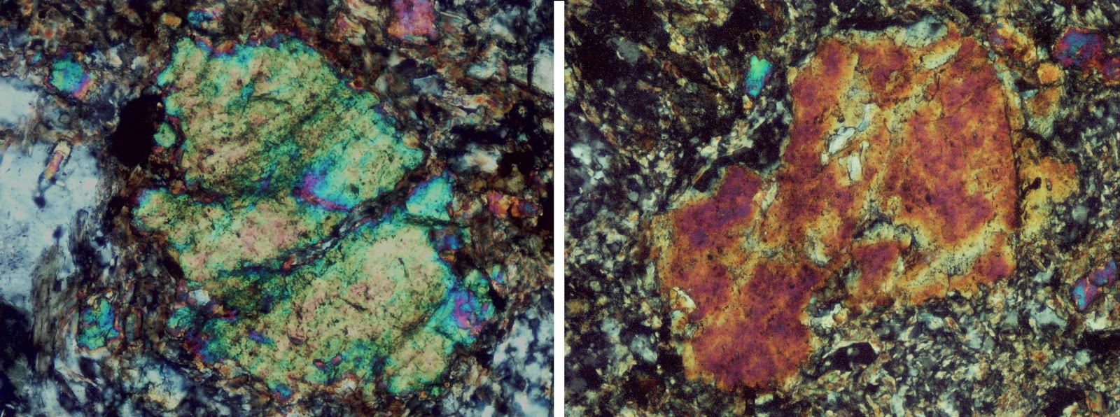
Abraded and corroded olivine (left) and augite (right) phenocrysts erupted in pyroclastic flow from Mt. Garibaldi, B. C., XPL. Each image covers 0.42 mm by 0.56 mm. Photos by Dan Snyder

Text

Abraded and embayed feldspar crystals with feldspar fragments altering to clay minerals or zeolites. Pyroclastic flow (tuff-breccia), Mt. Garibaldi, B. C., XPL.Imaged area 1.3 mm by 2 mm. Photo by Dan Snyder
Text

Feldspar fragments. Pyroclastic flow (tuff-breccia), Mt. Garibaldi, B. C., XPL.Imaged area 0.5 mm by 0.8 mm. Photo by Dan Snyder
 Text
 
Feldspar fragments in cloudy silica gel, altering to clay minerals or zeolites. Pyroclastic flow (tuff-breccia), Mt. Garibaldi, B.C., XPL. Imaged area 0.09 mm by 0.14 mm, 40X objective. Photo by Dan Snyder
Text

Feldspar fragments and devitrified volcanic glass. See next image for enlargement of area in rectangle. Pyroclastic flow (tuff-breccia), Mt. Garibaldi, B. C., XPL.Imaged area 0.5 mm by 0.8 mm. Photo by Dan Snyder
 Text


Text
Digitally-enlarged (6X) view of area outlined blue rectangle at upper right of previous image, showing fibrous fringe of neoforming mineral, likely a zeolite.
 Text


Text
Devitrified volcanic glass in tuff-breccia, Mt. Garibaldi, B.C., XPL. Imaged area 0.09 mm by 0.13 mm, 40X objective. Photo by Dan Snyder.
Text
Neoformed mineral mass, likely an iron-rich phyllosilicate, possibly nontronite, surrounding a relict mafic mineral crystal. Pyroclastic flow (tuff-breccia), Mt. Garbaldi, B. C. .XPL. Imaged area 0.5 mm by 0.8 mm. Photo by Dan Snyder.
Text
As above, ordinary light. Note opaque iron-oxide grains in former core of a mafic mineral in the center of  the brown-stained mineral mass.
 Text


Saturday, December 15, 2012

Eclogite from Norway - full thin section

Click on image to enlarge.          Specimen: Oliver Pluemper; Photo: Dan Snyder
Full thin section of eclogite from western Norway. Darkest (opaque) shapes are garnet; large brown mass at upper left is orthopyroxene; pale yellow and light brown grains at left and upper right are mostly clinopyroxene; gray, black and white grains at lower right and lower center are quartz. Macrophotograph in XPL. Imaged area 22mm by 43mm.

See post of June 14, 2012 for detailed description of rock and higher-magnification photomicrographs.

Many thanks to Oliver Pluemper of the University of Oslo for the specimen.

Tuesday, October 30, 2012

Groundmass minerals (replacing pyroxene) forming around well-defined nucleii in greenstone.


Click on image to enlarge.      © Daniel R. Snyder
Above: Neocrystallization of mesostasis (groundmass) minerals around well-defined nucleii.  Note how boundaries of replacement mineral grains iform a subtle mosaic pattern in the same area as the linear pattern of remaining pyroxene structure, which is accentuated by green stringers of chlorite. (center and right). Older mesostasis is on left. Greenstone no. 97, cobble from glacial gravel, southeastern Michigan, provenance likely northern Ontario. PPL. Imaged area 0.13 mm by 0.2 mm (40x objective)

Click on image to enlarge.      © Daniel R. Snyder

Above: XPL. Imaged area 0.5 mm by 0.8 mm. (10x objective). Remnants of pyroxene are yellow-green; altered pyroxene is light brown; dark brown and gray grains are mesostasis. Area covered by 40x image (top of this post) is outlined in white.


Click on image to enlarge.      © Daniel R. Snyder

Above: PPL. Imaged area 0.5 mm by 8 mm. (10x objective0. Area covered by 40x image (top of this post) is outlined in white.

 
Click on image to enlarge.      © Daniel R. Snyder

Above: PPL Imaged area 0.25 mm by 0.4 mm (20x objective). Only the upper three-quarters of the 40x image, outlined in white, is included in this view.


Sunday, October 28, 2012

Twinned Clinopyroxene in Greenstone

Click on image to enlarge.         © Daniel R. Snyder

Twinned clinopyroxene in greenstone no. 108, cobble from glacial gravel, southeastern Michigan, likely provenance northern Ontario. XPL. Imaged area 0.5 mm by 8 mm. (10x objective).

The extinction angle for this and similar crystals in the specimen is 42 degrees. A helpful viewer of this blog suggested that this might be pigeonite. However, after digging deeper into the characteristics of pyroxenes, I found that pigeonite doesn't occur in metamorphic rocks.

Here's an optical scan of a rough-polished section of the hand specimen. Imaged area 11 mm by 17 mm.


Texr


Wednesday, August 22, 2012

Fine-grained poikiloblastic greenstone


Click on image to enlarge.          Photo © Daniel R. Snyder
Optical scan of rough-polished section of fine-grained greenstone cobble. Throughout the specimen, larger grains enclose numerous smaller grains: poikiloblastic texture. Cobble from glacial gravel in southeastern Michigan, provenance likely northern Ontario. Imaged area 6.35 cm by 8.67 cm.


Click on image to enlarge.          Photo © Daniel R. Snyder
Higher-magnification view of an area near the center of the previous image, clearly showing the inclusions in almost all of the larger grains. Imaged area 10 mm by 17 mm.


Click on image to enlarge.          Photo © Daniel R. Snyder
Image illustrating the small grain size typical of this greenstone specimen. Enlarge this image to see the smaller individual grains in the darker areas. XPL, 10X objective. Imaged area 0.5 mm by 0.8 mm.

Click on image to enlarge.          Photo © Daniel R. Snyder
A plagioclase lath (center ) has been almost entirely replaced by epidote. XPL, 10X objective. Imaged area 0.5 mm by 0.8 mm.


Click on image to enlarge.          Photo © Daniel R. Snyder
 As above, PPL. Epidote is indicated by high-relief green areas.

Thursday, June 14, 2012

Eclogite from Norway


Click on image to enlarge.         Specimen: Oliver Pluemper; Photo: Dan Snyder

Eclogite hand specimen from Norway. Thanks to Oliver Pluemper, University of Oslo, for the specimen. Optical scan. Specimen width 9.42 cm, height 7.3 cm.

Click on image to enlarge.     Specimen: Oliver Pluemper     Photo: Dan Snyder
Garnet (large, dark forms) and pyroxene (small and very  small yellow  and brown forms) in eclogite specimen shown in hand sample image above. This specimen, from the western Gneiss belt of Norway, has undergone significant deviatoric stress, which the garnet has resisted more effectively than has the pyroxene. XPL. Imaged area 2.7 mm by 4 mm.

Monday, February 20, 2012

Eclogite pebble fragment

Click on image to enlarge.          Specimen: Michael Davis; Photo: Dan Snyder
A slice through a small eclogite pebble from San Onofre breccia. Red and brown garnet with green pyroxene. Thanks to Distinguished Professor Michael Davis, University of California - Riverside, for the specimen. Found as beach rock, Camp Pendleton (USMC), California. Optical scan of sawed surface. Long dimension of imaged area 33 mm.


Click on image to enlarge.          Photo: Dan Snyder
Reflected-light photomicrograph of a small portion of the above sample. 2x objective. Imaged area 2.7mm by 4mm.


Click on image to enlarge.          Photo: Dan Snyder
Higher-magnification image of left center of previous image. Subhedral pyroxene grains visible at right of large garnet grain. Black grains are rutile, a common accessory in eclogite. Reflected-light photomicrograph. 4x objective. Imaged area 1.3mm by 2mm.

Click on image to enlarge.         Photo: Dan Snyder
Photomicrograph of part of thin section of same specimen at same scale as previous image. Black forms are garnet grains, brightly colored grains are pyroxene. Note many inclusions in garnet. Beige triangular grain at right is amphibole. XPL. Imaged area 1.3mm by 2mm.