 |
| Click on image to enlarge. Photo © Daniel R. Snyder |
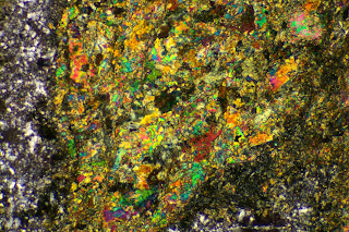
Metamorphosed orthopyroxene (left, bottom, upper right, lower right) and plagioclase (center) in norite. Pyroxene grains show varying degrees and types of metamorphism. Original linear structure of large pyroxene grain at left is detectable only as a weak lination imparted by dark specks. Plagioclase is more intact, but shows degradation (blurring) of twin lamellae. Mt. Prospect complex, Litchfield County, northwestern Connecticut. XPL. Imaged area 1.3mm x 2mm.
 |
| Click on image to enlarge. Photo © Daniel R. Snyder |
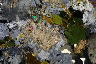
Higher-magnification image of replacement product (blue) in large orthopyroxene grain (left side of top photo). XPL. Imaged area 0.5mm by 0.8mm.
 |
| Click on image to enlarge. Photo © Daniel R. Snyder |
Higher-magnification (10x objective) image of heavily-altered orthopyroxene grain (lower right corner of top photo). Green areas are remnants of original mineral. Note linear (chain silicate) structure. Also note that small grains of opaque mineral, presumably an iron-oxide mineral, are preferentially concentrated in pyroxene remnants. XPL. Imaged area 0.5mm by 0.8mm.
 |
| Click on image to enlarge. Photo © Daniel R. Snyder |
PPL image of above, showing relict lineation and concentration of opaques in pyroxene remnants.
 |
| Click on image to enlarge. Photo © Daniel R. Snyder |
Higher-magnification (40x objective) image of cluster of opaques in center of photo above. Note that many of the "opaques" appear to be slightly translucent brown ovals, aligned to the lineation in the pyroxene. PPL. Imaged area 0.13mm by 0.2mm.


